Cell Types
The following cell types were identified in Cliona lampa as described by Rutzler and Rieger (1973).
o Forms the pinacoderm (exterior surface of the sponge). Lines the aquiferous system and
o Flat, squamous cells that are closely packed next to each other forming a sheet. Pinacocytes also line the channels that are created by the sponge as it bores through substrate.
o These cells were identified upon light microscope observations of cross sections of the Cliona sp. found on Heron Island (Below).
.jpg) 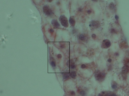 Left: Flat, squamous pinacocyte cell
Right: Enlarged pinacocyte (boxed)
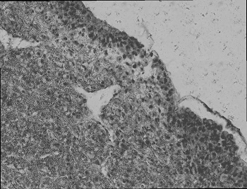 .jpg) Left: Image displaying tightly packed cells in pinacoderm (100x)
Left: Image displaying tightly packed cells in pinacoderm (100x)
Right: Enlarged pinacodermo Always associated with collagen
o Appear at transition from ectosome to choanosome and are very abundant in choanosome.
- Choanocytes:
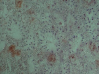
o Multiple choanocytes form a spherical shape with flagella pointing inwards. This is known as a choanocyte chamber.
o Important in water flow and feeding
o Found in Cliona sp. on Heron Island (Right: Image showing choanocyte chambers with dinstinct flagella pointing inwards).
o Abundant in burrowing process together with specialised cells known as etching cells (described below). Distinguished by large nucleus and round shape
o Totipotent cells: act like stem cells allowing process of regeneration. Can differentiate into any type of sponge cell (Ruppert et al. 2004).
o Macrophage-like: phagocytic and involved in digestion and internal transport.
o These cells were identified in the Cliona sp. found on Heron Island (show on image)
o Found throughout the choanosome.
o Secrete spicules (structural support for sponges).
o Possible spicules identified in Cliona sp. cross section on observation under a light microscope.
Image showing two possibe spicules (centre) in Cliona sp. cross section
o Etching cells are a type of archaeocyte that is the main cell involved in the pr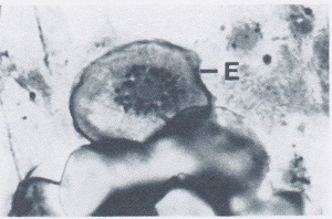 ocess of bioerosion. Either by chemical or mechanical means, these etching cells penetrate the substratum forming tunnels throughout the substrate. ocess of bioerosion. Either by chemical or mechanical means, these etching cells penetrate the substratum forming tunnels throughout the substrate.
o These cells are elongate with an orientation perpendicular to the substrate. The cell is widest at the distal end and is tapered towards base closest to the substratum to allow penetration (Cobb 1969).

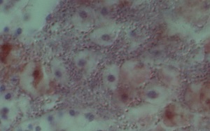 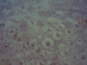
Comparison of etching cells
Top: Etching cell described by Cobb (1969)
Bottom: Etching cell found in Cliona sp. cross section.
Possible CaCO3 fragments were found associated with etching cells. As an H & E stain was used to stain the sponge material, a red colour is produced in the presence of a basic substance from the eosin and the acidic substances turn purple-blue from the haemotoxylin. As CaCO3 is organic material thus basic, this may explain the red colouration visible on the cross section as it is generally found over the etching cells.
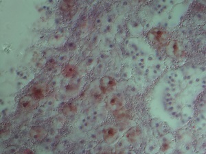 Image showing red colouring associated with etching cells possibly indicating
the presence of CaCO3 fragments as a result of its boring nature.
In this particular Cliona sp., it was noted that that cells are very small and compacted, therefore difficult to distinguish between different cell types on observation of the cross sections under a light microscope.
|